UltraScan Version

Manual


SOMO - Residue Definition Module:

SOMO Screen

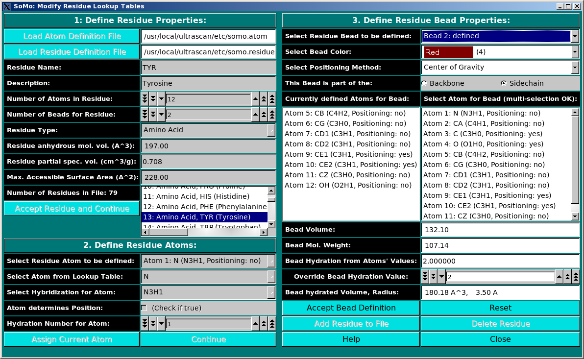
This module is used to define all residues that can be found in PDB files. In this module, you can also define the rules which are used to convert them into beads. You can add new residues or modify the properties of the existing ones. Three panels are available:

  1. Define Residue Properties
  2. Define Residue Atoms
  3. Define Residue Bead Properties

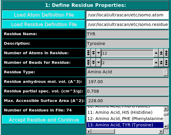
Panel 1: Define Residue Properties:

Define Residue Properties

In this panel, you can add or modify the general properties of a residue. 
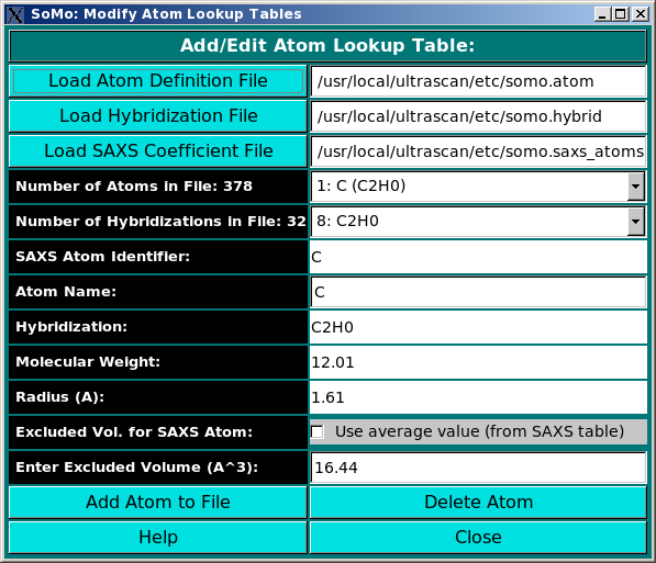
To add a new residue, you must first load the atom table containing the atomic groups, as defined in the PDB file (N, CA, CB, etc.), that make up the residue.
The residue name (ALA, GLY, PRO, etc.) is entered in the Residue Name field, with a description entered in the Description field (this will help in discriminating between entries having the same residue name in PDB files, but differing by the presence/absence of some atoms, like the PO2 group in nucleosides/nucleotides). The Number of Atoms in Residue field is then completed (note that only non-hydrogen atoms have to be counted; for NMR-derived models, hydrogen atoms fields are discarded by the SOMO PDB parser).
The next field, Number of Beads for Residue, defines how many beads will be used to represent the residue. For amino acids or nucleic acid bases, we suggest to use a bead for the main-chain and another for the side-chain segments, plus one for the PO2 group in nucleotides. For carbohydrates, a bead per residue will usually suffice. More complicated entries, like heme or NAD, must be assigned to a reasonable number of beads taking into account geometric and chemical considerations. Many entries are already defined in the somo.residue file distributed with the program.
The Residue Type field is then defined from the pull-down list, presently including:

  • Amino Acid
  • Sugar Moiety
  • Nucleotide
  • Heme
  • Phosphate
  • Co-factor
  • Ion
  • Detergent
  • Other

The Residue anhydrous mol. vol. (A^3) field has to be filled with a value usually derived from crystallography (see Tsai et al., J. Mol. Biol. 290:253-266, 1999; Nadassy et al., Nucl. Acid Res. 29:3362-3376, 2001; Voss and Gerstein, J. Mol. Biol. 346:477-492, 2005; Perkins, Eur. J. Biochem. 157:169-180, 1986). This is the volume that defines the total anhydrous volume of the bead(s) that will be used to model the residue, and for non tabulated entries it can be computed using on-line programs such as the 3V Contact Volume Calculator (http://www.molmovdb.org/cgi-bin/3v.cgi; a 0 Angstrom probe radius should be used). For ions, we used the values in Table I of Kiriukhin and Collins (Biophys. Chem. 99:155-168, 2002).
In contrast, the Residue partial spec. vol. (cm^3/g) field pertains to the solution partial specific volume (vbar) of the residue (in water @ 20°C), and is used to compute the global vbar of the model, necessary to determine the sedimentation coefficient from the translational frictional coefficient. Tabulated values are available for amino acids and some carbohydrates (see Perkins, Eur. J. Biochem. 157:169-180, 1986), or can be computed using the Cohn and Edsall method (Cohn and Edsall, Proteins, amino acids and peptides as ions and dipolar ions, Reinhold Publ. Comp., 1965, pp. 370-381; see also Durchschlag and Zipper, Biophys. Chem. 93: 141-157, 2001; Durchschlag and Zipper, J. Appl. Cryst. 30:803-807, 1997; Durchschlag and Zipper, Prog. Colloid Polym. Sci. 94:20-39, 1994). For ions, it was computed directly from the anhydrous mol. vol. (cm3) multiplied by Avogadro's number and divided by the molecular weight. Be aware, however, that there's no guarantee that a computed vbar will be correct: the use of an experimental vbar is strongly recommended!! (see also the vbar fields in the Miscellaneous Options editor.
Finally, the Max. Accessible Surface Area (A^2) field is entered. It is used for control purposes only, to calculate the percentage accessible surface area (ASA) of each residue with respect to this theoretical maximum value. For amino acids, it was taken from the tabulated values of Gly-XXX-Gly tripeptides (Miller et al., J. Mol. Biol. 196:641-656, 1987). For ions, it was computed from the radius (Table I of Kiriukhin and Collins, Biophys. Chem. 99:155-168, 2002). For the other entries currently present in the somo.residue file, it was computed on isolated moieties using the 3V Contact Volume Calculator (see above) with a 1.4 A probe radius.

When all fields have been filled, the residue is accepted by pressing the Accept Residue and Continue button, and the Number of Residues in File field is consequently updated.

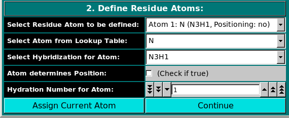
Panel 2: Define Residue Atoms:

Define Residue Atoms

In this panel, the atoms making up the residue are chosen, using the atom table previously selected. The atoms are numbered sequentially up to the value entered in the Number of Atoms in Residue field in the previous panel, and can be chosen from the Select Residue Atom to be defined pull-down menu.
An atom type is then selected from the pull-down menu in the Select Atom from Lookup Table menu, and an available hybridization is then chosen from the Select Hybridization for Atom pull-down menu below it.
If the current atom will contribute to the definition of the position of the bead to which it will be assigned (next panel), the Atom determines Position checkbox must be selected. Be aware that in the default mode operation, the SOMO method will place the amino acid main-chain bead using the "peptide bond rule", i.e., it will place the bead at the center of gravity between the (CA-C-O)n and the (N)n+1 atoms (special rules apply when PRO is the (n+1) residue). If the peptide bond rule is deselected in the Miscellaneous Options editor, then all the atom positioning rules defined in the somo.residue file will apply.
The Hydration Number for Atom field is used to assign an integer number of water molecules to the current atom. This field was recently introduced to allow for the direct hydration of a structure for SAXS computations. Currently, we have assigned directly to atoms only the amino acids hydration values, derived from the theoretical hydration numbers for entire residues of Kuntz and Kauzmann (Adv. Protein Chem. 28:239-345, 1974). The global hydration number for each bead in the residue is subsequently computed by summing up each atom's value. This calculated global value can be replaced with another value using the Override Bead Hydration Value button in the third panel of this editor (see below).
The Assign Current Atom button is then clicked, and the Select Residue Atom to be defined field is then updated.
This operation is repeated until all atoms in the residue have been assigned, and the Continue button can then be pressed.

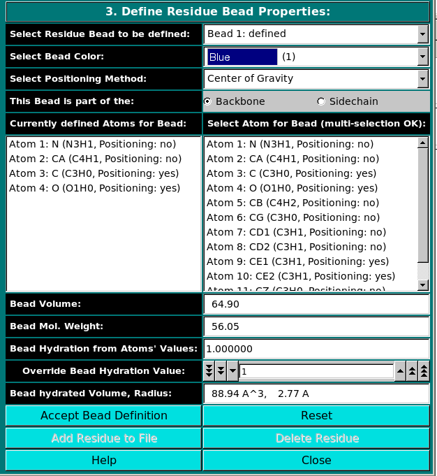
Panel 3: Define Residue Bead Properties:

Define Residue Bead Properties

In the last panel of this module, the atoms in each residue are assigned to bead(s), and each bead's properties are then defined.
According to the value entered in the Number of Beads for Residue field in Panel 1, the Select Residue Bead to be defined pull-down menu will allow to select a bead from the list. All subsequent operations will affect the selected bead only.
First a color is assigned to the bead using the Select Bead Color pull-down menu. Be aware that two colors are presently reserved to "label" the beads so that they can be automatically excluded from the hydrodynamic computations, colors "0" (black) and "6" (brown), so they should NOT be used. To avoid they inadvertent selection, a Warning pop-up will appear if color "0" or "6" are chosen, with this message: "Black and brown are reserved colors, please chose a different color". In addition, we color-code "7" (white) exposed beads that are fused together, and "8" (grey) beads that were originally labeled as buried ("6") but are found to be above the exposure threshold upon re-check. In the residue table (an excerpt of the current somo.residue is shown) provided with the UltraScan distribution, the colors are assigned so to label the beads as:

    0. - Black: RESERVED, automatically assigned to very small beads (always excluded from computations)
  1. - Blue: protein main-chain
  2. - Green: protein acidic side-chain (D,E)
  3. - Cyan: protein hydrophobic side-chain (A,V,L,I,F,W)
  4. - Red: protein polar side-chain (H,Y,S,T,N,Q)
  5. - Magenta: protein non-polar side-chain (C,M,P)
  6. - Brown: RESERVED for buried beads (automatically assigned during model generation)
  7. - White: USED for fused beads
  8. - Grey: USED for previously buried beads found to be exposed on re-check
  9. - Light Blue: lipids tails, carbon monoxide
  10. - Light Green: USED by the Automatic Bead Builder for non-coded residues (see here)
  11. - Light Cyan: bases in DNA/RNA, oxygen, 13P
  12. - Light Red: heme, NAD, other co-factors, some prosthetic groups and ions, PO2, lipids heads
  13. - Light Magenta: carbohydrates (including sugar rings in nucleotides), some ions
  14. - Yellow: protein basic side-chain (K,R)
  15. - Bright White: unassigned


Next comes the Select Positioning Method field, offering three options:

Center of gravity: between the atoms assigned to the bead and marked "yes" in the Atom determines Position checkbox in Panel 2;
Farthest atom: from the center of gravity of the molecule, again between the atoms assigned to the bead and marked "yes" in the Atom determines Position checkbox in Panel 2;
No positioning: to exclude the bead from the model.
Currently, only the Center of gravity option is being used.

Each bead needs then to be defined as belonging to the backbone or to the sidechain of the molecule by clicking the appropriate box in the This Bead is part of the: field. This determines at which sequential stage the bead will be processed during overlap removal in the SoMo method.

The next step is the assignment of atoms to the bead. In the Select Atom for Bead (multi-selection OK) window, you can see the list of all the atoms that belong to the residue, as defined previously in Panel 2. By clicking on an atom, it will be also displayed in the Currently defined Atoms for Bead window to the left. Once all atoms belonging to a bead have been selected, the bead is accepted by clicking on the Accept Bead Definition button.

The Bead Volume field defines the anhydrous volume (in A3) assigned to the bead. IMPORTANT: the sum of the volumes of the bead(s) defining a residue MUST match the Residue anhydrous mol. vol. entered in Panel 1. The program will NOT let you save the residue/bead definition if there is a discrepancy between these two values. The values present in the current definitions of the amino acid residues were derived from the crystallographic analysis (see above for references). When a molecule needs to be subdived in more than one bead, the volumes of the various parts can be determined using the 3V Contact Volume Calculator (see above). However, the parts' volumes will NOT add up to the entire molecule volume, and MUST be then proportionally rescaled. The volume of all other entries in the current somo.residue table were determined in this way.

The next field, Bead Mol. Weight is for visualization only, and will display the bead molecular weight, calculated from the values (taken from the atom table.

Likewise, the subsequent field, Bead Hydration from Atoms' Values, will show the theoretical number of water of hydration molecules assigned to the bead, based on the sum of the atoms' values entered in Panel 2. However, since the atomic values are likely not defined for most residues, a global value for each bead can be entered in the next field, Override Bead Hydration Value. Most beads' values currently present in the somo.residue file were derived from literature. For carbohydrates, we used the values of Shiio (J. Am. Chem. Soc. 80:70-73, 1958). For ions, we used the values in Table I of Kiriukhin and Collins (Biophys. Chem. 99:155-168, 2002). For the other residues, and for further information, see Brookes et al., Eur. Biophys. J. in press, 2009, and Rai et al., Structure 13:723-734, 2005. The volume of these water molecule(s) (defined under the Miscellaneous Options; see here) will be later automatically added to the anhydrous volume of the bead to define its hydrated volume and radius.

Finally, in the last field, Bead hydrated Volume, Radius, is again for visualization only, and will display the bead's hydrated molecular volume and hydrated radius (derived by summing to the anhydrous Bead Volume the volume of the water of hydration molecules, see above).

If the residue is represented by more than one bead, the process is repeated until all beads have been processed.

The residue can then be added to the file by clicking on the Add Residue to File button. The process can be interrupted at any stage by pressing the Reset button.

To Edit an already defined residue, double-click on its name on the list (that can be scrolled) present in the window next to the Number of residues in File field in Panel 1. All the stored properties will then appear first in the Panel 1 fields, which can then be updated. By clicking on the Accept Residue and Continue button, the properties listed in Panel 2 become then accessible, and can be likewise edited. Finally, the Panel 3 options will become again available for editing by clicking on the Continue button in Panel 2. Accept Bead Definition and Add Residue to File buttoms will then allow to update the residue table (somo.residue).
A selected residue/bead definition can be also removed from the file by clicking on the Delete Residue button.
Pressing Close will then exit from the Modify Residue Lookup Tables menu and return the operator to the Main menu.


www contact: Borries Demeler

This document is part of the UltraScan Software Documentation distribution.
Copyright © notice.

The latest version of this document can always be found at:

http://www.ultrascan.uthscsa.edu

Last modified on February 1, 2010.